X-ray photography of an oil painting
Analysis report
Inger Nyström Godfrey
Studio Västsvensk Konservering
VA 2019-00558
Date of report: 2019 07 18
All material in this report, both text and images, is published under the CC BY-ND license.

Introduction
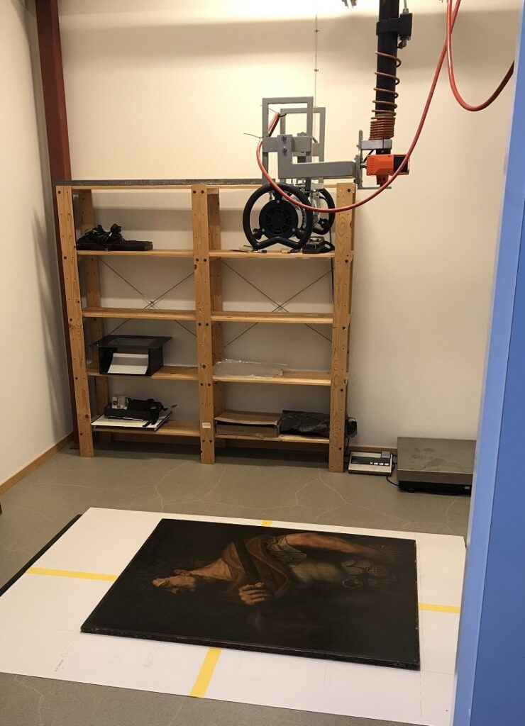
An x-ray examination of an older oil painting was carried out on the request of the owners. The images were delivered to the customer in DICOM format together with a light version of the x-ray software program that can read and manage these files. Also included are the original images in TIF format and a set of filtered images, which can be stitched together with image processing software.
Subject
The painting depicts the Roman emperor Domitian (Domitianus). According to information from the client, the picture of Domitian was the last in a series of 12 portraits of Roman emperors. The first eleven portraits are said to have been painted by Titian in the 1530s, while it is unclear who painted the twelfth portrait. The series is said to have been sold and transported to England from where it was later again sold to the Spanish Royal house. It is also said that in Spain the paintings were destroyed by fire during the 18th century. However, according to the client, there is uncertainty about this.
The now x-rayed painting of Domitian was for sale in the 1920s by an English auction house as a painting by Titian. The painting is lined and retouched, unclear when and by whom. A 14C dating has been performed at the Tandem Laboratory in Uppsala, Sweden, and a research engineer at the laboratory states: ”Unfortunately, the dating ends up in a non-linear area. The probability is higher that the canvas is from the late 15th century than the late 16th century, more than that we probably cannot say”.
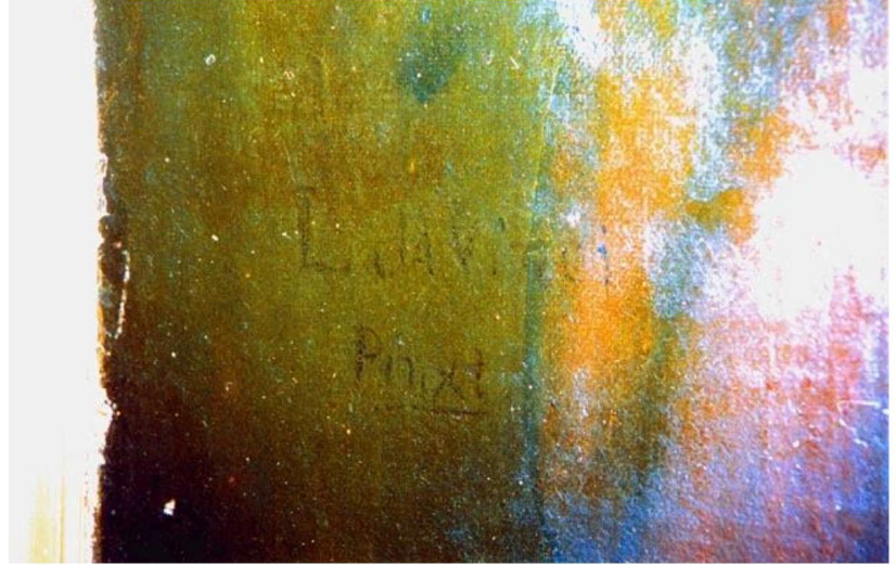
A faint signature ”L da Vinci, Pinxt (or Pnixt)” can be seen under the man’s right elbow (fig. 7). Depending on the light source, this is more or less visible.
Aim, method and questions
The X-ray analysis is carried out to see if there is another painting on the canvas under the visible one and/or if you can see if the artist (or someone else later) has made adjustments to the motif. The aim is also to find out if you can see the signature L da Vinci using X-rays.
Stitching the images into one complete image will be performed by the client.
Analysis
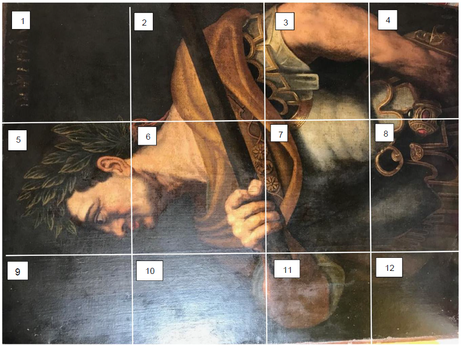
The X-ray analysis was performed with a digital industrial X-ray (CR). Exposure facts are presented in table 1. Twelve image plates covered the entire surface of the painting with some overlap and have been numbered 1-12, see figure 1. The lines drawn up on the photograph (fig 1) show the approximate position of the image plates. The reason why the squares look different in size is probably because the photo of the painting was not taken completely straight or vertical. A separate x-ray image was taken of the area where the signature is.
Radiation source; Sitex CPseries, type CP160D. Scanner: Carestream Industrex HPX-1. Imaging plate:
Carestream Industrex Flex XL Blue Digital Imaging Plate 5537.
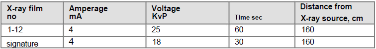
Table 1. Exposure facts for each X-ray film
Results and Observations
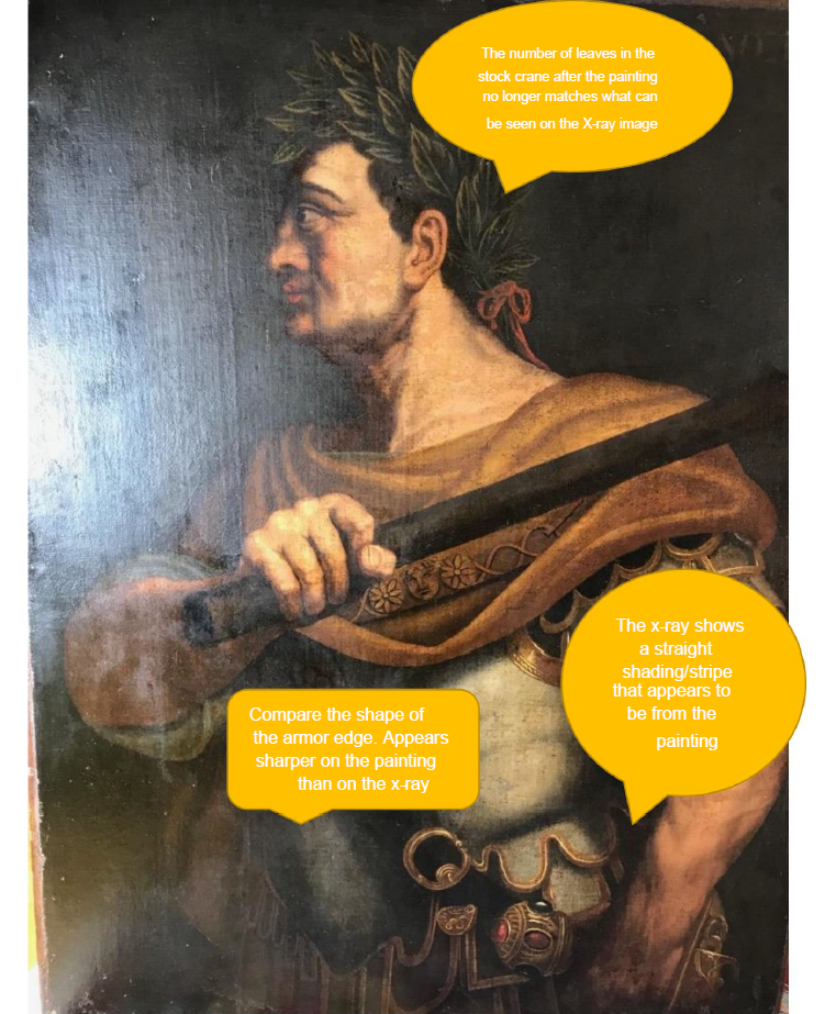
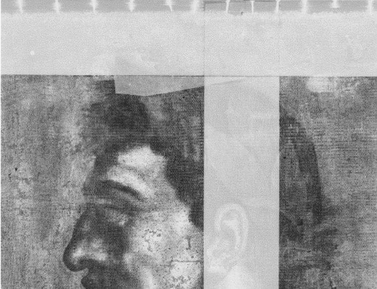
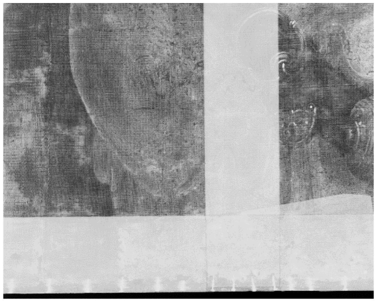
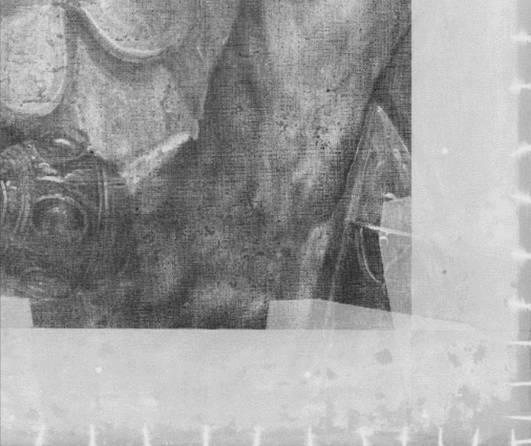
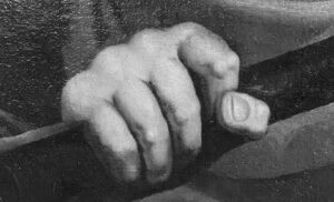
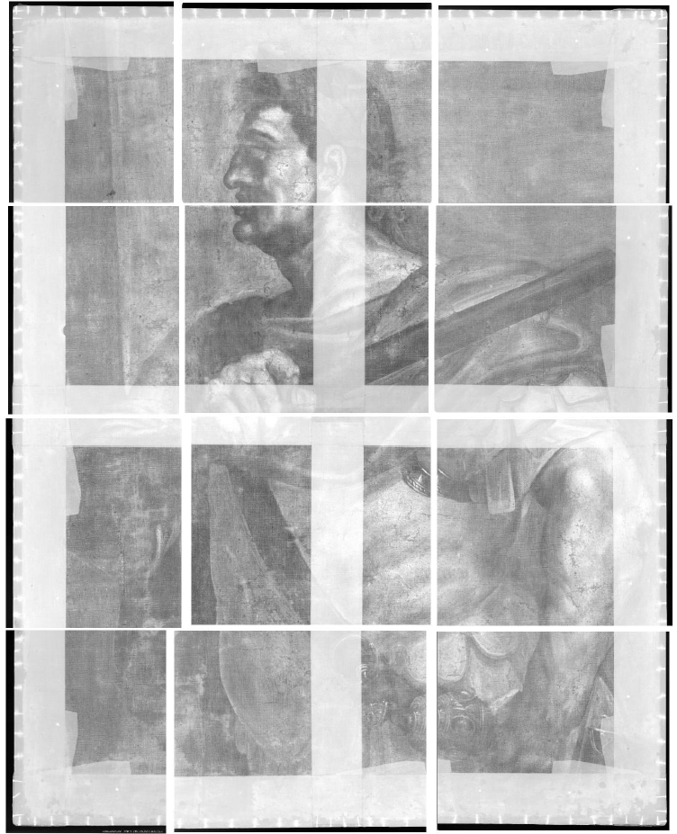
The motif, weave structure, stretcher, damages and brush strokes are clearly visible in the images, but no traces of any underlying painting could be seen on initial inspection, however, there are details on the painting that cannot be seen on the x-rays, indicating that it has been ”improved” on one or more occasions (fig. 3-6). The laurel wreath has more leaves on the painting than can be seen on the x-ray of the same area. The shape of the armor edge appears to be more pointed on the painting than on the x-ray image and finally on x-ray image 4 a faint straight shadow of something that does not appear to be on the painting is visible. These comparisons between the X-ray images and the painting were made using a photograph of the painting and not the painting itself, so there may be a possible source of error here. The x-ray images (in DICOM format) should of course be studied more carefully by an expert.
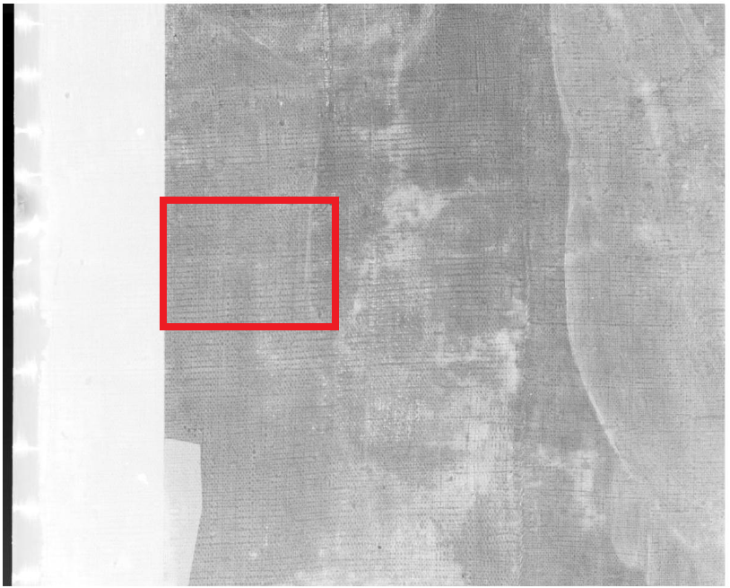
Figure 2 shows the x-ray images (TIF format) in the correct position, but not stitched together. Figures 7 and 8 show the area with the signature (photo from the client and x-ray). The signature was not made any clearer by using x-ray, in fact the signature could not be seen on the radiograph, which tells us that whatever pigment was used it was of low density and most likely of organic origin.
processed.